Biology & medicine · 1929
On the Antibacterial Action of Cultures of a Penicillium, with Special Reference to their Use in the Isolation of B. influenzæ
British Journal of Experimental Pathology · June 1929
- Source
- pmc.ncbi.nlm.nih.gov
- Retrieved
- 27 September 2026
- License
- Public domain
- Rights holder
- Alexander Fleming, 1929
- Language
- English
About this edition and its rights
Edition
The paper as printed in the British Journal of Experimental Pathology, vol. X, no. 3 (June 1929), pp. 226-236, with the two plates of Figs. 1 to 4, from the scan in PubMed Central (PMC2048009, file brjexppathol00255-0037.pdf, 13 pages at 600 ppi). Its text layer was corrected word by word against the page images, with the help of a second, independent OCR (the Internet Archive microfilm scan sim_international-journal-of-experimental-pathology_1929-06_10_3). The title, author and affiliation are set in the header of the poster; running heads, page numbers and the signature mark are left out; words hyphenated at line ends are joined (post-nasal keeps its hyphen, as in the title of Table IV); the spaces before ; and : are not kept. Spellings of the print are kept, among them influenzæ beside influenzae, diphtheriæ beside diphtheriae, “Pfeiffers bacillus”, “It doses not”, “Des moissures du group”, and “(Fildes, 1921)” in the text for the reference dated 1920. The tables are set as tables: the dashes that mark no growth in Tables I and IV are set as minus signs; the ditto marks are set as “,,”; the sign that marks uncountable colonies in the table of the rate of killing is set as ∝, the glyph of the print; the two halves of Table IV, printed side by side (cases 1 to 13 and 14 to 25), are set as one table; Table III, printed sideways on p. 230, is set upright. Tables II and III follow the paragraph that introduces them. The four photographs are the plates of the scan, cropped to each figure with its engraved labels, in grayscale; their captions are set as printed (“Fig. 4—” without a full stop). The short rule before the acknowledgement is kept.
Rights
Published in 1929, before 1931: public domain in the United States. Alexander Fleming, its only author, died on 11 March 1955, before 1956: public domain in France since 1 January 2026.
The PubMed Central record of the article (OAI-PMH front matter of PMC2048009) names Alexander Fleming as its only author and carries no copyright statement or license. A scan of a public domain page adds no protected work.
Received for publication May 10th, 1929.
While working with staphylococcus variants a number of culture-plates were set aside on the laboratory bench and examined from time to time. In the examinations these plates were necessarily exposed to the air and they became contaminated with various micro-organisms. It was noticed that around a large colony of a contaminating mould the staphylococcus colonies became transparent and were obviously undergoing lysis (see Fig. 1).
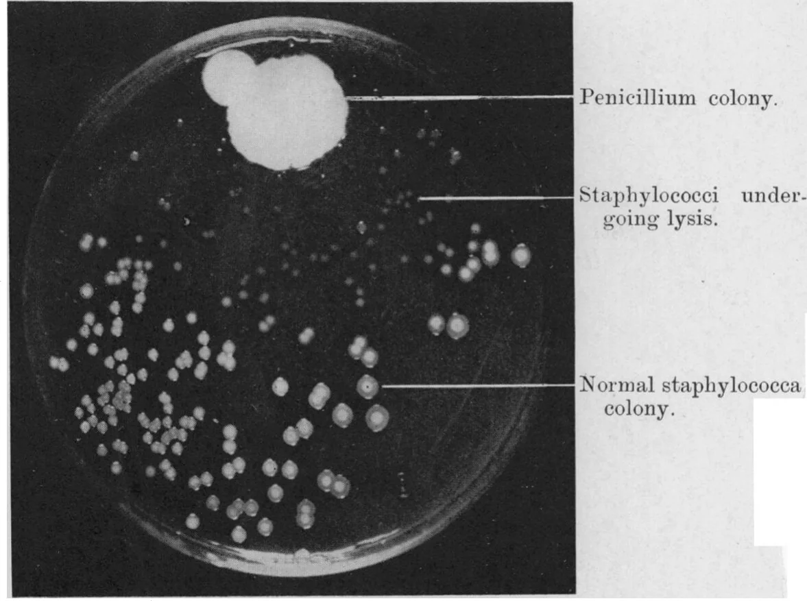
Subcultures of this mould were made and experiments conducted with a view to ascertaining something of the properties of the bacteriolytic substance which had evidently been formed in the mould culture and which had diffused into the surrounding medium. It was found that broth in which the mould had been grown at room temperature for one or two weeks had acquired marked inhibitory, bactericidal and bacteriolytic properties to many of the more common pathogenic bacteria.
CHARACTERS OF THE MOULD.#
The colony appears as a white fluffy mass which rapidly increases in size and after a few days sporulates, the centre becoming dark green and later in old cultures darkens to almost black. In four or five days a bright yellow colour is produced which diffuses into the medium. In certain conditions a reddish colour can be observed in the growth.
In broth the mould grows on the surface as a white fluffy growth changing in a few days to a dark green felted mass. The broth becomes bright yellow and this yellow pigment is not extracted by CHCl3. The reaction of the broth becomes markedly alkaline, the pH varying, from 8·5 to 9. Acid is produced in three or four days in glucose and saccharose broth. There is no acid production in 7 days in lactose, mannite or dulcite broth.
Growth is slow at 37°C. and is most rapid about 20°C. No growth is observed under anaerobic conditions.
In its morphology this organism is a penicillium and in all its characters it most closely resembles P. rubrum. Biourge (1923) states that he has never found P. rubrum in nature and that it is an “animal de laboratoire.” This penicillium is not uncommon in the air of the laboratory.
IS THE ANTIBACTERIAL BODY ELABORATED IN CULTURE BY ALL MOULDS?#
A number of other moulds were grown in broth at room temperature and the culture fluids were tested for antibacterial substances at various intervals up to one month. The species examined were: Eidamia viridiscens, Botrytis cineria, Aspergillus fumigatus, Sporotrichum, Cladosporium, Penicillium, 8 strains. Of these it was found that only one strain of penicillium produced any inhibitory substance, and that one had exactly the same cultural characters as the original one from the contaminated plate.
It is clear, therefore, that the production of this antibacterial substance is not common to all moulds or to all types of penicillium.
In the rest of this article allusion will constantly be made to experiments with filtrates of a broth culture of this mould, so for convenience and to avoid the repetition of the rather cumbersome phrase “Mould broth filtrate,” the name “penicillin” will be used. This will denote the filtrate of a broth culture of the particular penicillium with which we are concerned.
METHODS OF EXAMINING CULTURES FOR ANTIBACTERIAL SUBSTANCE.#
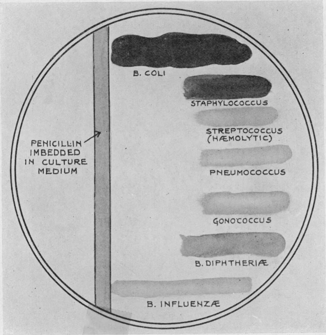
The simplest method of examining for inhibitory power is to cut a furrow in an agar plate (or a plate of other suitable culture material), and fill this in with a mixture of equal parts of agar and the broth in which the mould has grown. When this has solidified, cultures of various microbes can be streaked at right angles from the furrow to the edge of the plate. The inhibitory substance diffuses very rapidly in the agar, so that in the few hours before the microbes show visible growth it has spread out for a centimetre or more in sufficient concentration to inhibit growth of a sensitive microbe. On further incubation it will be seen that the proximal portion of the culture for perhaps one centimetre becomes transparent, and on examination of this portion of the culture it is found that practically all the microbes are dissolved, indicating that the anti-bacterial substance has continued to diffuse into the agar in sufficient concentration to induce dissolution of the bacteria. This simple method therefore suffices to demonstrate the bacterio-inhibitory and bacteriolytic properties of the mould culture, and also by the extent of the area of inhibition gives some measure of the sensitiveness of the particular microbe tested. Fig. 2 shows the degree of inhibition obtained with various microbes tested in this way.
The inhibitory power can be accurately titrated by making serial dilutions of penicillin in fresh nutrient broth, and then implanting all the tubes with the same volume of a bacterial suspension and incubating them. The inhibition can then readily be seen by noting the opacity of the broth.
For the estimation of the antibacterial power of a mould culture it is unnecessary to filter as the mould grows only slowly at 37° C., and in 24 hours, when the results are read, no growth of mould is perceptible. Staphylococcus is a very suitable microbe on which to test the broth as it is hardy, lives well in culture, grows rapidly, and is very sensitive to penicillin.
The bactericidal power can be tested in the same way except that at intervals measured quantities are explanted so that the number of surviving microbes can be estimated.
PROPERTIES OF THE ANTIBACTERIAL SUBSTANCE.#
Effect of heat.—Heating for 1 hour at 56° or 80° C. has no effect on the antibacterial power of penicillin. Boiling for a few minutes hardly affects it (see Table II). Boiling for 1 hour reduces it to less than one quarter its previous strength if the fluid is alkaline, but if it is neutral or very slightly acid then the reduction is much less. Autoclaving for 20 minutes at 115° C. practically destroys it.
Effect of filtration.—Passage through a Seitz filter does not diminish the antibacterial power. This is the best method of obtaining sterile active mould broth.
Solubility.—It is freely soluble in water and weak saline solutions. My colleague, Mr. Ridley, has found that if penicillin is evaporated at a low temperature to a sticky mass the active principle can be completely extracted by absolute alcohol. It is insoluble in ether or chloroform.
Rate of development of inhibitory substance in culture.—A 500 c.c. Erlenmeyer flask containing 200 c.c. of broth was planted with mould spores and incubated at room temperature (10° to 20° C.). The inhibitory power of the broth to staphylococcus was tested at intervals.
| After | 5 | days | complete | inhibition | in 1 in 20 | dilution. |
| ,, | 6 | ,, | ,, | ,, | ,, 1 in 40 | ,, |
| ,, | 7 | ,, | ,, | ,, | ,, 1 in 200 | ,, |
| ,, | 8 | ,, | ,, | ,, | ,, 1 in 500 | ,, |
Grown at 20° C. the development of the active principle is more rapid and a good sample will completely inhibit staphylococci in a 1 in 500 or 1 in 800 dilution in 6 or 7 days. As the culture ages the antibacterial power falls and may in 14 days at 20° C. have almost disappeared.
The antibacterial power of penicillin falls when it is kept at room temperature. The rate of this fall can be seen from Table I.
| Growth of staphylococcus in dilutions of penicillin as under. | ||||||||||||
|---|---|---|---|---|---|---|---|---|---|---|---|---|
| 1/20. | 1/40. | 1/60. | 1/80. | 1/100. | 1/200. | 1/300. | 1/400. | 1/600. | 1/800. | 1/1000. | Control. | |
| At time of filtration | − | − | − | − | − | − | − | − | − | ± | ++ | ++ |
| After 4 days | − | − | − | − | − | − | − | − | − | ± | ++ | ++ |
| ,, 7 ,, | − | − | − | − | − | − | − | ± | + | + | ++ | ++ |
| ,, 9 ,, | − | − | − | − | − | − | − | ± | + | + | ++ | ++ |
| ,, 13 ,, | − | − | − | − | − | + | + | + | + | + | ++ | ++ |
| ,, 15 ,, | − | ± | + | + | + | + | + | + | + | + | ++ | ++ |
If the reaction of penicillin is altered from its original pH of 9 to a pH of 6·8 it is much more stable.
The small drops of bright yellow fluid which collect on the surface of the mould may have a high antibacterial titre. One specimen of such fluid completely inhibited the growth of staphylococci in a dilution of 1 in 20,000 while the broth in which the mould was growing, tested at the same time, inhibited staphylococcal growth in 1 in 800.
If the mould is grown on solid medium and the felted mass picked off and extracted in normal salt solution for 24 hours it is found that the extract has bacteriolytic properties.
If this extract is mixed with a thick suspension of staphylococcus suspension and incubated for 2 hours at 45° C. it will be found that the opacity of the suspension has markedly diminished and after 24 hours the previously opaque suspension will have become almost clear.
Influence of the medium on the antibacterial titre of the mould culture.—So far as has been ascertained nutrient broth is the most suitable medium for the production of penicillin. The addition of glucose or saccharose, which are fermented by the mould with the production of acid, delays or prevents the appearance of the antibacterial substance. Dilution of the broth with water delays the formation of the antibacterial substance and diminishes the concentration which is ultimately reached.
INHIBITORY POWER OF PENICILLIN ON THE GROWTH OF BACTERIA.#
Tables II and III show the extent to which various microbes, pathogenic and non-pathogenic, are inhibited by penicillin. The first table shows the inhibition by the agar plate method and the second shows the inhibitory power when diluted in nutrient broth.
| Dilution of penicillin in broth. | ||||||||||||
|---|---|---|---|---|---|---|---|---|---|---|---|---|
| 1/5. | 1/10. | 1/20. | 1/40. | 1/80. | 1/100. | 1/200. | 1/400. | 1/800. | 1/1600. | 1/3200. | Control. | |
| Staphylococcus aureus | 0 | 0 | 0 | 0 | 0 | 0 | 0 | 0 | ± | ++ | ++ | ++ |
| ,, epidermidis | 0 | 0 | 0 | 0 | 0 | 0 | 0 | 0 | ± | ++ | ++ | ++ |
| Pneumococcus | 0 | 0 | 0 | 0 | 0 | 0 | 0 | 0 | 0 | ++ | ++ | ++ |
| Streptococcus (hæmolytic) | 0 | 0 | 0 | 0 | 0 | 0 | 0 | 0 | 0 | ± | ++ | ++ |
| ,, viridans (mouth) | 0 | 0 | 0 | 0 | 0 | 0 | ± | ++ | ++ | ++ | ++ | ++ |
| ,, fæcalis | ++ | ++ | ++ | ++ | ++ | ++ | ++ | ++ | ++ | ++ | ++ | ++ |
| B. anthracis | 0 | 0 | + | + | ++ | ++ | ++ | ++ | ++ | ++ | ++ | ++ |
| B. pseudo-tuberculosis rodentium | + | + | ++ | ++ | ++ | ++ | ++ | ++ | ++ | ++ | ++ | ++ |
| B. pullorum | + | + | ++ | ++ | ++ | ++ | ++ | ++ | ++ | ++ | ++ | ++ |
| B. dysenteriæ | + | ++ | ++ | ++ | ++ | ++ | ++ | ++ | ++ | ++ | ++ | ++ |
| B. coli | ++ | ++ | ++ | ... | ... | ... | ... | ... | ... | ... | ... | ++ |
| B. typhosus | ++ | ++ | ++ | ... | ... | ... | ... | ... | ... | ... | ... | ++ |
| B. pyocyaneus | ++ | ++ | ++ | ... | ... | ... | ... | ... | ... | ... | ... | ++ |
| B. proteus | ++ | ++ | ++ | ... | ... | ... | ... | ... | ... | ... | ... | ++ |
| V. choleræ | ++ | ++ | ++ | ... | ... | ... | ... | ... | ... | ... | ... | ++ |
| 1/60. | 1/120. | 1/300. | 1/600. | Control. | ||||||||
| B. diphtheriæ (3 strains) | 0 | ± | ++ | ++ | ++ | |||||||
| Streptococcus pyogenes (13 strains) | 0 | 0 | 0 | ++ | ++ | |||||||
| ,, ,, (1 ,, ) | 0 | 0 | ± | ++ | ++ | |||||||
| ,, fæcalis (11 ,, ) | ++ | ++ | ++ | ++ | ++ | |||||||
| ,, viridans at random from fæces (1 strain) | 0 | 0 | 0 | ++ | ++ | |||||||
| ,, ,, ,, ,, (2 strains) | 0 | 0 | ± | ++ | ++ | |||||||
| ,, ,, ,, ,, (1 strain) | 0 | ± | ++ | ++ | ++ | |||||||
| ,, ,, ,, ,, (1 ,, ) | + | ++ | ++ | ++ | ++ | |||||||
| ,, ,, ,, ,, (1 ,, ) | ++ | ++ | ++ | ++ | ++ | |||||||
| ,, ,, at random from mouth (1 ,, ) | 0 | ± | ++ | ++ | ++ | |||||||
| ,, ,, ,, ,, (2 strains) | 0 | 0 | ++ | ++ | ++ | |||||||
| ,, ,, ,, ,, (1 strain) | 0 | 0 | 0 | ++ | ++ | |||||||
| 0 = no growth; ± = trace of growth; + = poor growth; ++ = normal growth. | ||||||||||||
| Type of microbe. | Extent of inhibition in mm. from penicillin embedded in agar, serum agar, or blood agar plates. | |
|---|---|---|
| Unheated. | Boiled for 1 minute. | |
| Experiment 1: | ||
| Staphylococcus pyogenes | 23 | 21 |
| Streptococcus ,, | 17 | 17 |
| ,, viridans (mouth) | 17 | 15 |
| Diphtheroid bacillus | 27 | 22 |
| Sarcina | 10 | 10 |
| Micrococcus lysodeikticus | 6 | 7 |
| ,, from air (1) | 20 | 16 |
| ,, ,, (2) | 4 | 9 |
| B. anthracis | 0 | 0 |
| B. typhosus | 0 | 0 |
| Enterococcus | 0 | 0 |
| Experiment 2: | ||
| Staphylococcus pyogenes | 24 | ... |
| Streptococcus ,, | 30 | ... |
| ,, viridans (mouth) | 25 | ... |
| Pneumococcus | 30 | ... |
| Diphtheroid bacillus | 35 | ... |
| B. pyocyaneus | 0 | ... |
| B. pneumoniæ (Friedlander) | 0 | ... |
| B. coli | 0 | ... |
| B. paratyphosus A | 0 | ... |
| Experiment 3: | ||
| Staphylococcus pyogenes | 16 | ... |
| Gonococcus | 16 | ... |
| Meningococcus | 17 | ... |
| Experiment 4: | ||
| Staphylococcus pyogenes | 17 | ... |
| ,, epidermidis | 18 | ... |
| Streptococcus pyogenes | 15 | ... |
| ,, viridans (fæces) | 5 | ... |
| B. diphtheriæ (2 strains) | 14 | ... |
| Diphtheroid bacillus | 10 | ... |
| Gram-negative coccus from the mouth (1) | 12 | .. |
| ,, ,, ,, (2) | 0 | ... |
| B. coli | 0 | ... |
| B. influenzæ (Pfeiffer) 6 strains | 0 | .. |
Certain interesting facts emerge from these Tables. It is clear that penicillin contains bacterio-inhibitory substance which is very active towards some microbes while not affecting others. The members of the coli-typhoid group are unaffected as are other intestinal bacilli such as B. pyocyaneus, B. proteus and V. cholerae. Other bacteria which are insensitive to penicillin are the enterococcus, some of the Gram-negative cocci of the mouth, Friedländer’s pneumobacillus, and B. influenzae (Pfeiffer), while the action on B. dysenteriae (Flexner), and B. pseudo-tuberculosis rodentium is almost negligible. The anthrax bacillus is completely inhibited in a 1 in 10 dilution but in this case the inhibitory influence is trifling when compared with the effect on the pyogenic cocci.
It is on the pyogenic cocci and on bacilli of the diphtheria group that the action is most manifest.
Staphylococci are very sensitive, and the inhibitory effect is practically the same on all strains, whatever the colour or type of the staphylococcus.
Streptococcus pyogenes is also very sensitive. There were small differences in the titre with different strains, but it may be said generally that it is slightly more sensitive than staphylococcus.
Pneumococci are equally sensitive with Streptococcus pyogenes.
The green streptococci vary very considerably, a few strains being almost unaffected while others are as sensitive as S. pyogenes. Gonococci, meningococci, and some of the Gram-negative cocci found in nasal catarrhal conditions are about as sensitive as are staphylococci. Many of the Gram-negative cocci found in the mouth and throat are, however, quite insensitive.
B. diphtheriæ is less affected than staphylococcus but is yet completely inhibited by a 1% dilution of a fair sample of penicillin.
It may be noted here that penicillin, which is strongly inhibitory to many bacteria, does not inhibit the growth of the original penicillium which was used in its preparation.
The Rate of Killing of Staphylococci by Penicillin.#
Some bactericidal agents like the hypochlorites are extremely rapid in their action, others like flavine or novarsenobillon are slow. Experiments were made to find into which category penicillin fell.
To 1 c.c. volumes of dilutions in broth of penicillin were added 10 c.mm. volumes of a 1 in 1000 dilution of a staphylococcus broth culture. The tubes were then incubated at 37°C. and at intervals 10 c.mm. volumes were removed and plated with the following result:
| Number of colonies developing after sojourn in penicillin in concentrations as under: | |||||
|---|---|---|---|---|---|
| Control. | 1/80. | 1/40. | 1/20. | 1/10. | |
| Before | 27 | 27 | 27 | 27 | 27 |
| After 2 hours | 116 | 73 | 51 | 48 | 23 |
| ,, 4½ ,, | ∝ | 13 | 1 | 2 | 5 |
| ,, 8 ,, | ∝ | 0 | 0 | 0 | 0 |
| ,, 12 ,, | ∝ | 0 | 0 | 0 | 0 |
It appears, therefore, that penicillin belongs to the group of slow acting antiseptics, and the staphylococci are only completely killed after an interval of over 4½ hours even in a concentration 30 or 40 times stronger than is necessary to inhibit completely the culture in broth. In the weaker concentrations it will be seen that at first there is growth of the staphylococci and only after some hours are the cocci killed off. The same thing can be seen if a series of dilutions of penicillin in broth are heavily infected with staphylococcus and incubated. If the cultures are examined after four hours it may be seen that growth has taken place apparently equally in all the tubes but when examined after being incubated overnight, the tubes containing penicillin in concentrations greater than 1 in 300 or 1 in 400 are perfectly clear while the control tube shows a heavy growth. This is a clear illustration of the bacteriolytic action of penicillin.
TOXICITY OF PENICILLIN.#
The toxicity to animals of powerfully antibacterial mould broth filtrates appears to be very low. Twenty c.c. injected intravenously into a rabbit were not more toxic than the same quantity of broth. Half a c.c. injected intraperitoneally into a mouse weighing about 20 gm. induced no toxic symptoms. Constant irrigation of large infected surfaces in man was not accompanied by any toxic symptoms, while irrigation of the human conjunctiva every hour for a day had no irritant effect.
In vitro penicillin which completely inhibits the growth of staphylococci in a dilution of 1 in 600 does not interfere with leucocytic function to a greater extent than does ordinary broth.
USE OF PENICILLIN TO DEMONSTRATE OTHER BACTERIAL INHIBITIONS.#
When materials like saliva or sputum are plated it is not uncommon to see, where the implant is thick, an almost pure culture of streptococci and pneumococci, and where the implant is thinner and the streptococcal colonies are more widely separated, other colonies appear, especially those of Gram-negative cocci. These Gram-negative cocci are inhibited by the streptococci (probably by the peroxide they produce in their growth) and it is only when the mass effect of the streptococci is reduced that they appear in the culture.
Penicillin may be used to give a striking demonstration of this inhibition of bacteria by streptococci and pneumococci. Sputum is spread thickly on a culture plate, and then 5 or 6 drops of penicillin is spread over one half of it. After incubation it may be seen that on the half untreated with penicillin there is a confluent growth of streptococci and pneumococci and nothing else, while on the penicillin-treated half many Gram-negative cocci appear which were inhibited by the streptococci and pneumococci, and can only flourish when these are themselves inhibited by the penicillin.
If some active penicillin is embedded in a streak across an agar plate planted with saliva an interesting growth sometimes results. On the portion most distal from the penicillin there are many streptococci, but these are obscured by coarsely growing cocci, so that the resultant growth is a copious confluent rough mass. These coarse growing cocci are extremely penicillin sensitive and stop growing about 25 mm. from the embedded penicillin. Then there is a zone of about 1 cm. wide of pure streptococci, then they are inhibited by the penicillin, and as soon as that happens Gram-negative cocci appear and grow right up to the embedded penicillin. The three zones of growth produced in this way are very striking.
USE OF PENICILLIN IN THE ISOLATION OF B. INFLUENZÆ (PFEIFFER) AND OTHER ORGANISMS.#
It sometimes happens that in the human body a pathogenic microbe may be difficult to isolate because it occurs in association with others which grow more profusely and which mask it. If in such a case the first microbe is insensitive to penicillin and the obscuring microbes are sensitive, then by the use of this substance these latter can be inhibited while the former are allowed to develop normally. Such an example occurs in the body, certainly with B. influenzæ (Pfeiffer) and probably with Bordet’s whooping-cough bacillus and other organisms. Pfeiffer’s bacillus, occurring as it does in the respiratory tract, is usually associated with streptococci, pneumococci, staphylococci and Gram-negative cocci. All of these, with the exception of some of the Gram-negative cocci, are highly sensitive to penicillin and by the addition of some of this to the medium they can be completely inhibited while B. influenzæ is unaffected. A definite quantity of the penicillin may be incorporated with the molten culture medium before the plates are made, but an easier and very satisfactory method is to spread the infected material, sputum, nasal mucus, etc., on the plate in the usual way and then over one half of the plate spread 2 to 6 drops (according to potency) of the penicillin. This small amount of fluid soaks into the agar and after cultivation for 24 hours it will be found that the half of the plate without the penicillin will show the normal growth while on the penicillin treated half there will be nothing but B. influenzæ with Gram-negative cocci and occasionally some other microbe. This makes it infinitely easier to isolate these penicillin-insensitive organisms, and repeatedly
B. influenzæ has been isolated in this way when they have not been seen in films of sputum and when it has not been possible to detect them in plates not treated with penicillin. Of course if this method is adopted then a medium favourable for the growth of B. influenzæ must be used, e. g. boiled blood agar, as by the repression of the pneumococci and the staphylococci the symbiotic effect of these, so familiar in cultures of sputum on blood agar, is lost and if blood agar alone is used the colonies of B. influenzæ may be so minute as to be easily missed.
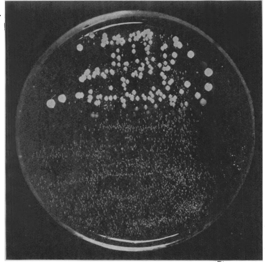
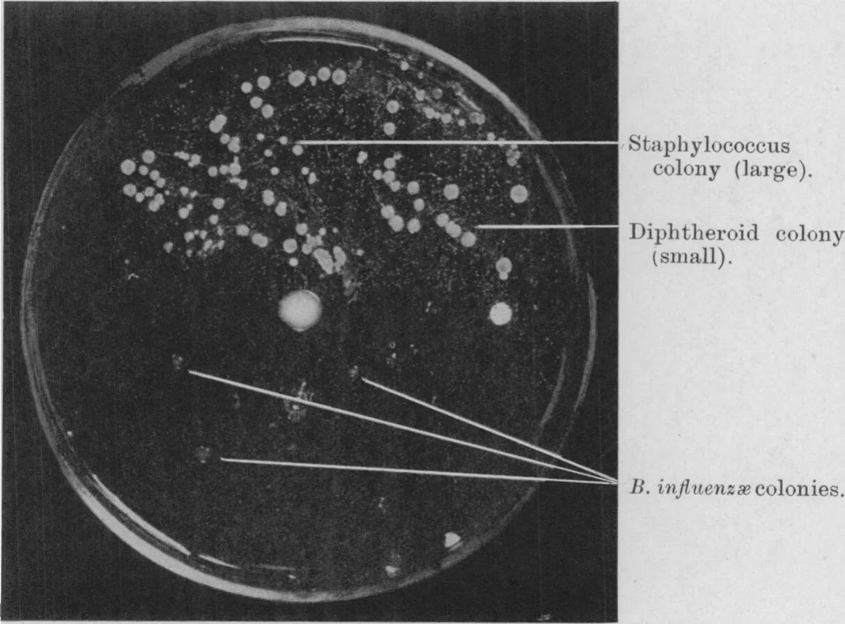
Figs. 3 and 4 are photographs of culture-plates made after the method described above. On the plate shown in Fig. 3 a mixture of staphylococci and B. influenzæ was spread over the whole plate of Fildes medium (Fildes, 1921), then 6 drops of penicillin were spread over the lower half of the plate. The upper half shows the mixed culture while the lower half gives a pure culture of B. influenzæ. Fig. 4 represents a culture of nasal mucus from a “cold” made on the same medium. Here, on the upper half (untreated with penicillin) staphylococci and diphtheroid bacilli grow abundantly, while on the treated (lower) half only some three or four colonies of B. influenzæ appear.
In conjunction with my colleague, Dr. McLean, a series of cultures were made from the throats of 25 nurses warded for “influenza.” The swabs were planted on boiled blood agar and over half of each plate was spread 3 or 4 drops of penicillin. The results are set forth in Table IV.
| Without penicillin. | With penicillin. | |||||
|---|---|---|---|---|---|---|
| Pneumococcus or Streptococcus. | B. influenzæ. | Gram-negative cocci. | Pneumococcus or Streptococcus. | B. influenzæ. | Gram-negative cocci. | |
| 1. | ++ | + | + | − | ++ | + |
| 2. | ++ | ++ | ++ | − | ++ | + |
| 3. | ++ | ++ | + | − | + | − |
| 4. | + | − | − | − | + | + |
| 5. | ++ | − | − | − | ++ | − |
| 6. | ++ | − | − | − | ++ | ++ |
| 7. | ++ | − | ++ | − | + | + |
| 8. | + | + | − | − | + | − |
| 9. | ++ | − | − | − | + | + |
| 10. | ++ | − | − | − | + | − |
| 11. | ++ | − | − | − | + | − |
| 12. | ++ | + | ++ | − | + | + |
| 13. | + | ++ | ++ | − | + | ++ |
| 14. | ++ | − | − | − | + | − |
| 15. | ++ | − | − | − | + | + |
| 16. | ++ | − | − | − | + | + |
| 17. | ++ | − | − | − | + | + |
| 18. | ++ | − | − | − | + | + |
| 19. | ++ | + | − | − | + | ++ |
| 20. | ++ | − | − | − | − | + |
| 21. | ++ | − | − | − | + | + |
| 22. | ++ | − | − | − | + | − |
| 23. | + | − | + | − | + | + |
| 24. | ++ | ++ | − | − | ++ | − |
| 25. | ++ | − | ++ | − | − | − |
In the above Table account has only been taken of the common microbes found in these cultures. In some there were a few diphtheroid bacilli which were always penicillin sensitive, and in others there were Gram-negative bacilli which were penicillin insensitive, although they were inhibited by streptococci or pneumococci. Pneumococci and streptococci were classed together, as complete tests were not made to differentiate one from the other.
(From the appearance of the colonies and the morphological characters pneumococci were evidently present in most cases in much larger numbers than were streptococci.)
The swabs were generally planted thickly and in some cases where the growth on the portion of the plate without penicillin was almost confluent, the cultures were sampled by taking smears from thick portions of the growth. In these cases it is possible that the results given do not give a quite complete picture of the cultures. This, however, does not affect the present argument that by the addition of penicillin to the culture medium, and the consequent inhibition of the pyogenic cocci, the isolation of B. influenzæ is very much easier. And in a number of cases it was isolated when it was completely missed in the cultures without penicillin.
It is quite immaterial how many pneumococci and streptococci are present in a specimen—they are completely inhibited—and even a few B. influenzæ can be isolated from a mixture with an enormous number of these cocci.
From a number of observations which have been made on sputum, post-nasal and throat swabs it seems likely that by the use of penicillin, organisms of the B. influenzæ group will be isolated from a great variety of pathological conditions as well as from individuals who are apparently healthy.
DISCUSSION.#
It has been demonstrated that a species of penicillium produces in culture a very powerful antibacterial substance which affects different bacteria in different degrees. Speaking generally it may be said that the least sensitive bacteria are the Gram-negative bacilli, and the most susceptible are the pyogenic cocci. Inhibitory substances have been described in old cultures of many organisms; generally the inhibition is more or less specific to the microbe which has been used for the culture, and the inhibitory substances are seldom strong enough to withstand even slight dilution with fresh nutrient material. Penicillin is not inhibitory to the original penicillium used in its preparation.
Emmerich and other workers have shown that old cultures of B. pyocyaneus acquire a marked bacteriolytic power. The bacteriolytic agent, pyocyanase, possesses properties similar to penicillin in that its heat resistance is the same and it exists in the filtrate of a fluid culture. It resembles penicillin also in that it acts only on certain microbes. It differs however in being relatively extremely weak in its action and in acting on quite different types of bacteria. The bacilli of anthrax, diphtheria, cholera and typhoid are those most sensitive to pyocyanase, while the pyogenic cocci are unaffected, but the percentages of pyocyaneus filtrate necessary for the inhibition of these organisms was 40, 33, 40 and 60 respectively (Bocchia, 1909). This degree of inhibition is hardly comparable with 0·2% or less of penicillin which is necessary to completely inhibit the pyogenic cocci or the 1% necessary for B. diphtheriæ.
Penicillin, in regard to infections with sensitive microbes, appears to have some advantages over the well-known chemical antiseptics. A good sample will completely inhibit staphylococci, Streptococcus pyogenes and pneumococcus in a dilution of 1 in 800. It is therefore a more powerful inhibitory agent than is carbolic acid and it can be applied to an infected surface undiluted as it is non-irritant and non-toxic. If applied, therefore, on a dressing, it will still be effective even when diluted 800 times which is more than can be said of the chemical antiseptics in use. Experiments in connection with its value in the treatment of pyogenic infections are in progress.
In addition to its possible use in the treatment of bacterial infections penicillin is certainly useful to the bacteriologist for its power of inhibiting unwanted microbes in bacterial cultures so that penicillin insensitive bacteria can readily be isolated. A notable instance of this is the very easy isolation of Pfeiffers bacillus of influenza when penicillin is used.
In conclusion my thanks are due to my colleagues, Mr. Ridley and Mr. Craddock, for their help in carrying out some of the experiments described in this paper, and to our mycologist, Mr. la Touche, for his suggestions as to the identity of the penicillium.
SUMMARY.#
- A certain type of penicillium produces in culture a powerful antibacterial substance. The antibacterial power of the culture reaches its maximum in about 7 days at 20° C. and after 10 days diminishes until it has almost disappeared in 4 weeks.
- The best medium found for the production of the antibacterial substance has been ordinary nutrient broth.
- The active agent is readily filterable and the name “penicillin” has been given to filtrates of broth cultures of the mould.
- Penicillin loses most of its power after 10 to 14 days at room temperature but can be preserved longer by neutralization.
- The active agent is not destroyed by boiling for a few minutes but in alkaline solution boiling for 1 hour markedly reduces the power. Autoclaving for 20 minutes at 115° C. practically destroys it. It is soluble in alcohol but insoluble in ether or chloroform.
- The action is very marked on the pyogenic cocci and the diphtheria group of bacilli. Many bacteria are quite insensitive, e.g. the coli-typhoid group, the influenza-bacillus group, and the enterococcus.
- Penicillin is non-toxic to animals in enormous doses and is non-irritant. It doses not interfere with leucocytic function to a greater degree than does ordinary broth.
- It is suggested that it may be an efficient antiseptic for application to, or injection into, areas infected with penicillin-sensitive microbes.
- The use of penicillin on culture plates renders obvious many bacterial inhibitions which are not very evident in ordinary cultures.
- Its value as an aid to the isolation of B. influenzæ has been demonstrated.
REFERENCES.#
Biourge.—(1923) ‘Des moissures du group Penicillium Link.’ Louvain, p. 172.
Emmerich, Loeuw and Korschun.—(1902) Zbl. Bakt., 30, 1.
Bocchia.—(1909) Ibid., 50, 220.
Fildes, P.—(1920) Brit. J. Exp. Path., 1, 129.